
Research
X-ray diffraction with high spatial resolution
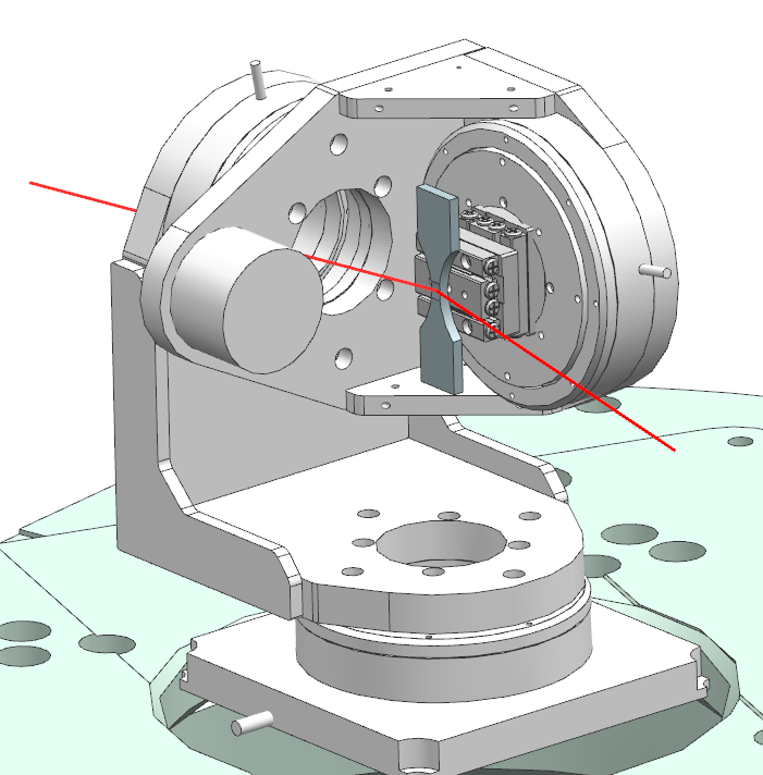 |
X-ray diffraction (XRD) is one of the most important and versatile techniques for determining the structure of materials from micrometer down to sub-nanometer scales. Taking advantage of the crystal-like structure XRD can accurtately measure microscopic sample features such residual stress, dislocation density or grain sizes, which control macroscopic properties like ductility, durability or the electronic band structure. In collaboration with the P06 beamline of PETRA III we are developing an approach to combine these capabilites with spatial resolutions down to micrometers and large penetration depths in metal samples. Together with our collaborators from the Material Systems for Lightweight Vehicle Construction group we will use this novel technique to get a deeper understanding of the impact of material properties on the micrometer scale on fatigue mechanisms. |
X-ray scattering for materials characterisation
|
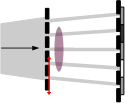
X-ray imaging based on the edge-illumination principle provides an X-ray scattering signal, which originates from sample features that may be considerably smaller than a detector pixel. Thus, edge-illumination is a scattering technique that truely allows for sub-pixel imaging, i.e., sensitivity towards micrometer and sub-micrometer features with pixels sizes of tens of micrometers and larger. Collaborating with the groups for High Frequency and Quantum Electronics and Forming technologies we want to investigate the application of X-ray scattering to technical composite materials in order to understand the origin and grow mechanism of defects during manufactoring. |
 |
X-ray fluorescence tomography of biological and nanoscale samples

|
X-ray fluorescence (XRF) is a technique that utilises x-ray induced secondary emission from atoms in order to achieve elemental and chemical sensitivity. Combining XRF with tomography as well as cutting-edge x-ray focussing optics individual elements can be mapped in 3D with spatial resolutions down to sub-micrometers. This is especially benefitial for the characterisation of biological samples as the elemental distribution is often a crucial clue for resolving biomedical questions. We are using XRF in order to characterise atherosclerotic plaques for the future preventation of strokes and to determine the 3D distribution of nanoparticles in cancer cells. |